Center for Brain Biology and Behavior
Scott Schrage, September 25, 2023
fMRI scans may improve diagnosis of sports-related concussion
VARIABILITY IN BRAIN FUNCTION EMERGES AS PROMISING BIOMARKER
The scrum leaves a black eye, the ruck an aching shoulder, the tackle a bruised leg.
But the concussion? It leaves a question mark.
“Concussion is a complex and difficult injury to diagnose because of the variability of its symptoms,” said Aron Barbey, who directs the Center for Brain, Biology and Behavior at the University of Nebraska–Lincoln.
Yet a recent Barbey-led study has found that the same variability responsible for confusing the diagnosis of sports-related concussion may also help clarify it.
The research showed that measuring the variability of oxygen delivery in the brain — differences detectable with functional MRI, or fMRI — can outperform a common neurocognitive test when it comes to identifying athletes who recently, or even repeatedly, experienced a concussion. Supplementing the usual assessments of memory, processing speed and reaction time with fMRI measures could improve the likelihood that athletes return to play only when they’re ready, Barbey said.
“Biomarkers derived from brain imaging may provide a more objective way to diagnose concussions, which would help to ensure that athletes and other individuals who have sustained a concussion receive the appropriate medical care,” said Barbey, the Mildred Francis Thompson Professor of psychology at Nebraska. “Many concussion symptoms are subjective, including difficulty concentrating, irritability and mood changes. These symptoms can only be reported by the person who is experiencing them, which can make it difficult for clinicians to assess the severity of the injury and track the progress of recovery.”
As part of the study, 50 athletes from English and Welsh rugby leagues — 29 recently diagnosed with a concussion, six diagnosed with multiple, and 15 who had not recently experienced one — completed an assessment that has emerged as a go-to for detecting mild traumatic brain injury. Known as Immediate Post-concussion Assessment and Cognitive Testing, or ImPACT, the assessment asks about potential concussion symptoms before tasking its takers with recalling or matching words, symbols, colors, letters and the like.
The athletes also underwent fMRI, a neuroimaging technique that measures blood flow as a proxy for brain activity. Neurons use oxygen when firing. In response, more blood — carrying the nanoscopic oxygen tanks known as hemoglobins — will flow to those active brain regions. Because hemoglobins feature different magnetic properties while holding oxygen than after releasing it, magnetism-sensitive MRI scanners can generate imagery of which brain regions are most active. Using fMRI, Barbey’s team derived a novel measure of signal variability to assess how concussion affects communication and connectivity among the brain’s networks.
Barbey and colleagues then used their ImPACT and fMRI data to train a machine-learning model aimed at correctly slotting each of the 50 participants into one of three categories: no concussion, concussion or repeat concussion. A model that was trained on variations in blood oxygen levels identified the concussed and repetitively concussed athletes substantially more often than did one trained on ImPACT data, which struggled to differentiate among the three groups. But a model trained on both blood oxygen variability and ImPACT data outperformed models relying on either source alone, correctly pegging 31 of the 50 participants.
Another, more surprising result could eventually help distinguish between athletes who recently experienced a single concussion and those who have suffered multiple. The variability of blood oxygen levels registered lower in single-concussion athletes, the team found, when compared against that of healthy participants. That lower variability seems to reflect a well-established increase in the functional coupling, or hyperconnectivity, of brain regions following concussion.
In athletes recovering from multiple concussions, Barbey’s team found that the opposite was true: Their blood oxygen variability came out markedly higher than the control group’s. That divergence speaks to the possibility that repeat concussions act on the brain in ways distinct from a single injury — potentially by curbing network communication and connectivity over time — and might help explain why multiple concussions pose a greater health risk.
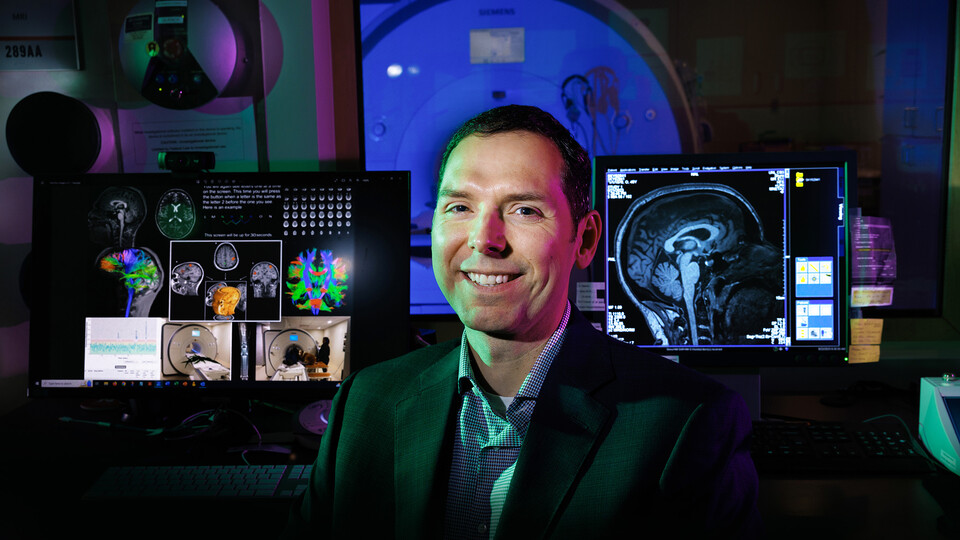
For now, Barbey said the study demonstrates that brain-imaging technology — including the MRI housed at the Center for Brain, Biology and Behavior, or CB3 — will play a central role in advancing the diagnosis of concussion. That’s especially heartening given that, though a few blood-based biomarkers have shown promise for detecting concussion, “there currently does not exist a gold-standard diagnostic test,” he said.
“Based on our findings and emerging evidence in the field, it’s clear that supplementing standard clinical tools with brain-imaging data adds value by improving the efficacy of concussion diagnosis,” Barbey said. “This suggests that we are indeed moving in the right direction to better understand and diagnose this complex injury.”
The chance to transfer that value from the southeast corner of Memorial Stadium, where CB3 resides, to the student-athletes who eat, train and play within it is among the reasons that Barbey recently moved to Lincoln and assumed leadership of the center.
“At CB3, we are inspired by the potential to develop new and more effective ways to diagnose and treat concussions,” he said. “This could have a major impact on the health and safety of our student-athletes, and it’s something that our Nebraska community is deeply committed to.”
The team reported its findings in the journal Brain Communications. Barbey authored the study with the University of Illinois Urbana-Champaign’s Evan Anderson, Tanveer Talukdar, Grace Goodwin, Valentina Di Pietro and Christopher Zwilling, along with the University of Birmingham’s Antonio Belli, David Davies and Kamal Yakoub. The researchers received funding in part from the National Institute for Health Research.


