Special Education and Communication Disorders
Chuck Green, December 4, 2019
Brain imaging helps bring cochlear implantation success into focus
For someone with hearing loss, a successful cochlear implant can change their world.
But because results vary among implant recipients, it is crucial to determine strong candidates for whom an implant will likely be successful.
A cochlear implant is a complex electronic device that can effectively restore hearing in individuals with severe to profound hearing loss. While the implant does not restore normal hearing and differs from hearing aids, which amplify sounds, it does provide a useful representation of sounds by directly stimulating the auditory nerve. The implant’s success depends on how well the auditory nerve functions.
Yingying Wang, assistant professor of special education and communication disorders, is collaborating with the University of Nebraska Medical Center on a three-year study designed to identify factors to determine the best candidates for successful cochlear implant procedures, and to explore relationships between the brain and speech perception outcomes — the process by which language is heard, interpreted and understood.
“We want to identify brain factors to better understand and predict potential for future hearing improvements among CI candidates,” said Wang, the project’s principal investigator.
Central to Wang’s research is learning how brain reorganization occurs before and after cochlear implantation. Brain images will be used to identify pre-surgical neural predictors that may provide a more comprehensive understanding of individual variability in cochlear implant outcomes, and therefore generate a more reliably informed prognosis.
Researchers plan to recruit 16 adults with post-lingual, severe-to-profound sensorineural hearing loss who are candidates for cochlear implantation.
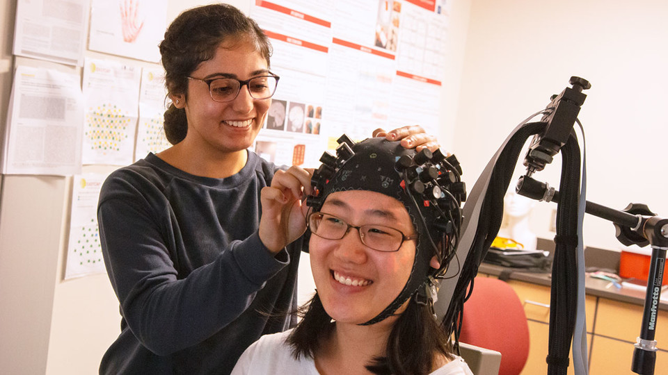
Before the surgical procedure, brain activity is measured by functional magnetic resonance imaging, and functional near-infrared spectroscopy and diffusion-weighted imaging measures the brain’s white matter integrity. This can reveal the initial functional areas in the brain responding to speech sounds and whether the auditory nerve is intact before surgery.
After implantation, only functional near-infared spectroscopy will be used to measure changes in brain activity together with changes in speech perception at three- and six-month follow-up visits. Post-implantation brain activity may clarify why some recipients have better speech perception.
The implantation procedure typically requires a one-day hospital stay to monitor potential complications. Recipients must also return a month after surgery for cochlear implant activation, and adjustments to its frequency and range to each individual.
“It’s not like getting a pair of eyeglasses,” Wang said. “There is a recovery and rehabilitation period for the brain to adapt to the implant.”
Wang is also interested in the brain’s neuroplasticity — its ability to change.
“We want to see how the brain adapts,” she said. “When someone has profound hearing loss, they have limited auditory stimulation. After the implant, they will have a lot of stimulation. We are interested to learn how the brain adapts to that.”
Since 1984, cochlear implants have benefited almost 325,000 individuals worldwide who are deaf or severely hard of hearing. There are approximately 96,000 users in the United States.
Although cochlear implants can significantly enhance auditory speech perception, the success rate differs widely across users. Variability factors include the patient’s age, duration of deafness, level of preoperative usable hearing and pre-implant hearing aid use.
However, such factors only explain only about 20% of the variance.
Wang said that because the procedure is invasive and costly, patients need a good understanding of what to expect, rather than simply receiving the cochlear implant and hoping for the best.
“Sometimes, patients with high expectations are disappointed with the results,” she said. “Some even want the implant removed.”
Wang’s findings may help doctors form a better prognosis for cochlear implant candidates, which will enable the patient to decide whether an implant is beneficial enough for them to proceed, or whether they should consider alternative options.
So far, Wang’s biggest challenge has been finding participants from Nebraska’s small pool of pre-surgical cochlear implant candidates. Johnathan Hatch, an otolaryngologist and assistant professor at UNMC who performs the procedure, has worked with area Veterans Affairs hospitals to recruit participants who experienced hearing damage during their military service.
Michelle Hughes, associate professor of special education and communication disorders, and Josh Sevier, assistant professor of practice, also have actively referred patients to Wang’s study.
The study is funded by the National Institute on Deafness and Other Communication Disorders and is housed at the Nebraska Center for Research on Children, Youth, Families and Schools and the Center for Brain, Biology and Behavior. Along with Wang, Hatch and Hughes, the research team also includes Hongying (Daisy) Dai, UNMC associate professor, who serves at the project’s statistician.
For more information on how to become a study participant, visit the Wang Lab website.


